| INTEGRA™
Bilayer Matrix Wound Dressing is placed on
a surgically debrided or excised wound, it provides
the needed framework for the blood vessels and dermal
skin cells to remodel the damaged site.
As skin cells migrate into the
matrix, the collagen is slowly absorbed and replaced
with collagen produced from the person's own cells.
In approximately 14 to 21 days, the scaffold is
eventually remodeled as the patient's cells rebuild
the damaged site and the silicone layer removed.
Complete wound closure occurs as epidermal cells
migrate from the wound edges. For larger wounds,
a thin skin graft of the person's epidermis may
be applied to the wound area to facilitate complete
wound closure.
Wound closure is typically
complete within 30 days. A person is left with a
healed wound created from their own tissue.
Clinical Sequence
|
DAY 0:
PRE-TREATMENT
All chronic wound patients
must have accurate diagnosis and treatment
of underlying disease and risks. There must
be thorough pre-operative control of inflammation,
ulceration, debris and bioburden, and edema
(as best as the disease and available treatments
permit).
|
 |
|
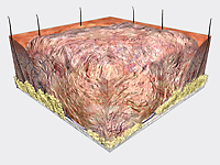
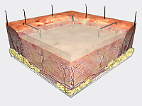
DAY 1:
DEBRIDEMENT
Prepare wound bed using
standard methods to ensure wound is free of
debris and necrotic tissue. Regardless of
how well the wound has been prepared and how
healthy it looks; INTEGRA™
Bilayer Matrix Wound Dressing must
not be placed on an existing wound surface.
The entire existing wound must be completely
excised or surgically debrided to ensure the
wound bed and edges contain viable tissue.
|
 |
|
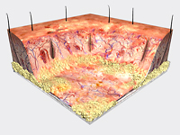
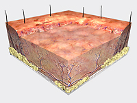
DAY 1:
APPLICATION
INTEGRA™ Bilayer Matrix
Wound Dressing is applied to the excised wound
bed. Fluids invade the matrix within minutes
of application, adhering it to the wound.
The Integra must conform to and contact the
wound surface. Tension within the material
will shear the matrix from the silicone, so
the material must not be stretched. It can
be affixed with sutures, staples, or any suitable
alternative.
|
 |
|
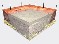
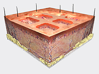
DAY 7-14:
CELLULAR INVASION and CAPILLARY GROWTH
Dermal cells begin migrating
into the matrix and establish a new vascular
network. The scaffold is eventually remodeled
as the patient's cells rebuild the damaged
site.
|
 |
|
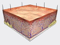
DAY 21+:
SILICONE REMOVAL
The silicone layer is removed.
The collagen template biodegrades and is absorbed
into the body.
|
 |
|
DAY 21-56+:
WOUND CLOSURE
Epidermal cells migrate
from the wound edges to complete wound closure.
For larger wounds, a thin epidermal autograft
may be considered to facilitate wound closure.
A thin 0.004 – 0.006 in. (0.1016 - 0.1524
mm) epidermal autograft may be applied over
the new remodeled skin.
Epidermal coverage over the wound yields a permanant
and lasting wound closure. |
 |
 |
|