|
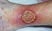
A 61-year-old woman had left leg
ulceration of many years duration and a history
of multiple venous thrombosis, pulmonary embolism,
and warfarin resistance. These are hallmark features
of a hypercoagulable disorder, but long standing
warfarin therapy precluded making the exact pre-thrombotic
diagnosis. In spite of the history, the usual stigmata
of venous disease, pigment, edema, and dermatosclerosis,
were not very severe, and there was never any improvement
by compression and topical care alone. This was
a hypercoagulable rather than a venous ulcer, further
confirmed histologically by microthrombi. Granulation
tissue at the base of the wound indicated intrinsic
wound healing competence, but there was chronic
active dermatitis, panniculitis, and necrosis in
the margins and periwound soft tissues.
|
 |


|
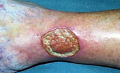
After a few weeks of hygiene,
good topical care, compression, and increased warfarin,
the wound and periwound were improved, but nevertheless,
inflammation and active necrosis-ulceration persisted
at the margins.
|
 |
|
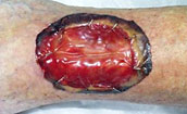
In surgery, the ulcer was excised,
including a tangential fibular ostectomy for hyperplastic
osteophytes (common under chronic inflammatory ulcers,
due to the effects of transforming or pro-proliferative
growth factors which are perpetually in the wound).
The wound was closed with Integra. Seen here at
6 days, periwound inflammation, erythema, and edema
have completely subsided.
|
 |
|
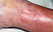
The ulcer remained healed during
a 4-year follow-up period, seen here 9 months after
skin grafts.
|
 |
Cases Courtesy of:
Marc E. Gottlieb, M.D., Jennifer Furman
Journal of Burns and Wounds, Vol 3, #2